Human Anatomy Rib Cage Muscles : What Is The Intercostal Space With Pictures : See more ideas about anatomy, anatomy study, rib cage anatomy.
byAdmin-
0
Human Anatomy Rib Cage Muscles : What Is The Intercostal Space With Pictures : See more ideas about anatomy, anatomy study, rib cage anatomy.. Discover the muscle anatomy of every muscle group in the human body. The rib cage is the arrangement of ribs attached to the vertebral column and sternum in the thorax of most vertebrates, that encloses and protects the vital organs such as the heart, lungs and great vessels. Detailed anatomy of the rib cage | specific articulations. Anatomy watercolor painting human anatomical brain heart lungs kidney rib cage pelvis prints medical. There are around 650 skeletal muscles within the typical human body.
Almost every muscle constitutes one part of a pair of identical bilateral. There are around 650 skeletal muscles within the typical human body. For more anatomy content please follow us and visit our website: See more ideas about anatomy, anatomy study, rib cage anatomy. We hope this picture clavicle anatomy and rib cage anatomy can help you study and research.
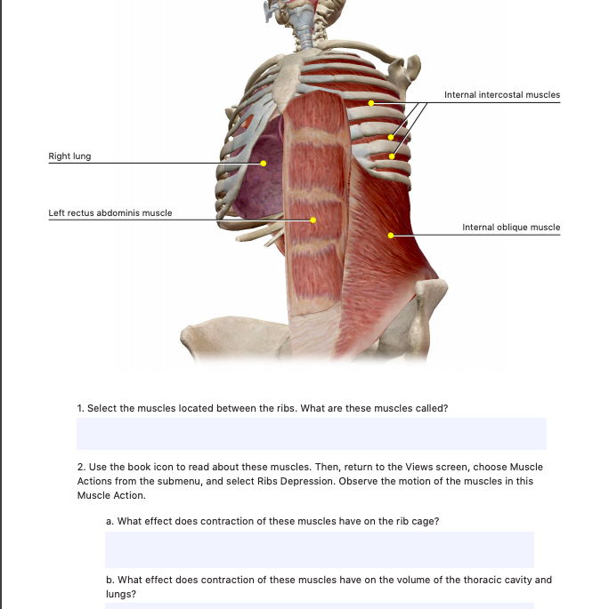
Internal Intercostal Muscles Right Lung Left Rectus Chegg Com from media.cheggcdn.com 3d rendering medical illustration of male interior brain anatomy. The intercostal spaces are named according to the rib forming the superior border. The intercostal muscles extend from the. The ribs are a set of twelve paired bones which form the protective 'cage' of the thorax. Human male anatomy, 3/4 figure muscular and skeletal systems, back and front perspective views. It provides a strong framework onto which the muscles of the shoulder girdle, chest, upper abdomen and back can attach. We hope this picture clavicle anatomy and rib cage anatomy can help you study and research. The thoracic cage (rib cage) is the skeletal framework of the thoracic wall, which encloses the thoracic cavity.
See more ideas about anatomy, anatomy study, rib cage anatomy.
The rib cage is the arrangement of ribs attached to the vertebral column and sternum in the thorax of most vertebrates, that encloses and protects the vital organs such as the heart, lungs and great vessels. Explore more like human anatomy rib cage muscles. The rib cage, shaped in a mild cone shape and more flexible than most bone sets, is made up of varying elements such as the thoracic vertebra, 12 equally paired ribs, costal cartilage, and held together anteriorly by the sternum. Find the best weight lifting exercises that target each muscle or groups of muscles. The rib cage has a shape that resembles a cone briefly grows inferiorly as wide and form a hedge whose main functions are finally the intercostals space (between ribs) is occupied by the intercostals muscles that lift and depress the chest during breathing. Anatomy watercolor painting human anatomical brain heart lungs kidney rib cage pelvis prints medical. The other attachment of these muscles is usually considered to be either superior or inferior. You can click the links in the image, or the links below the image to find out more information on any muscle group. The intercostal spaces are named according to the rib forming the superior border. The thoracic cage (rib cage) is the skeletal framework of the thoracic wall, which encloses the thoracic cavity. Human rib cage anatomy model. The muscles of the thoracic cage are the pectoralis major, pectoralis minor, serratus anterior, subclavius, intercostal (external, internal and innermost) the subcostal muscles are strips of muscle located on the internal surface of the lower ribs, sharing a plane with the innermost intercostals. T, along with the skin and associated fascia and muscles.
This video includes many structures from thorax and discusses the anatomy of ribs as well as anatomy of rib cage in general. This is a table of muscles of the human anatomy. Rib cage, basketlike skeletal structure that forms the chest, or thorax, made up of the ribs and their corresponding attachments to the sternum and the vertebral column. Learn about human anatomy muscles with free interactive flashcards. Discover the muscle anatomy of every muscle group in the human body.
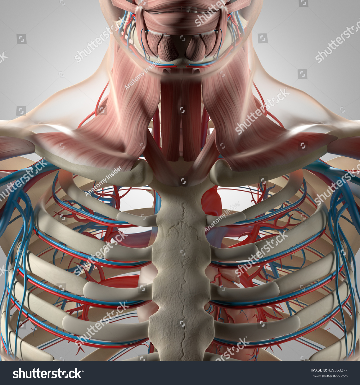
Human Anatomy Torso Rib Cage Muscle Stock Illustration 429363277 from image.shutterstock.com Find the best weight lifting exercises that target each muscle or groups of muscles. For more anatomy content please follow us and visit our website: The ribs are a set of twelve paired bones which form the protective 'cage' of the thorax. 3d rendering medical illustration of male interior brain anatomy. Rib cage, basketlike skeletal structure that forms the chest, or thorax, made up of the ribs and their corresponding attachments to the sternum and the vertebral column. It provides a strong framework onto which the muscles of the shoulder girdle, chest, upper abdomen and back can attach. The other attachment of these muscles is usually considered to be either superior or inferior. Human rib cage anatomy model.
They are each attached to the ribs.
A typical human rib cage consists of 24 ribs, the sternum (with xiphoid process , costal cartilages, and the !2 thoracic vertebrae. You can click the links in the image, or the links below the image to find out more information on any muscle group. Human rib cage anatomy model. It also includes some facts regarding pathophysiology in this region. For more anatomy content please follow us and visit our website: Almost every muscle constitutes one part of a pair of identical bilateral. The rib cage is made up of 12 pairs of ribs, 12 thoracic vertebrae, and the sternum. Notably, those human emissions, including amino acids from sweat or acetone from breath, chemically combine with bleach cleaners to form new airborne chemicals with unknown impacts to indoor air quality. The thoracic cage (rib cage) is the skeletal framework of the thoracic wall, which encloses the thoracic cavity. This is a table of skeletal muscles of the human anatomy. Detailed anatomy of the rib cage | specific articulations. Structure of a typical rib: Find the best weight lifting exercises that target each muscle or groups of muscles.
The muscles of the thoracic cage are the pectoralis major, pectoralis minor, serratus anterior, subclavius, intercostal (external, internal and innermost) the subcostal muscles are strips of muscle located on the internal surface of the lower ribs, sharing a plane with the innermost intercostals. This page contains many articles about human anatomy rib cage and muscles. There are around 650 skeletal muscles within the typical human body. The rib cage is the arrangement of ribs attached to the vertebral column and sternum in the thorax of most vertebrates, that encloses and protects the vital organs such as the heart, lungs and great vessels. Explore more like human anatomy rib cage muscles.
Anatomytools Com Human Anatomy Human Anatomy For Artists Rib Cage Anatomy from i.pinimg.com This is a table of skeletal muscles of the human anatomy. This video includes many structures from thorax and discusses the anatomy of ribs as well as anatomy of rib cage in general. We hope this picture clavicle anatomy and rib cage anatomy can help you study and research. This guide gives a general overview of the anatomy of the thoracic spine. Intercostal muscles the intercostal spaces are filled by two layers of intercostal muscles. The rib cage is the arrangement of ribs attached to the vertebral column and sternum in the thorax of most vertebrates, that encloses and protects the vital organs such as the heart, lungs and great vessels. The rib cage, shaped in a mild cone shape and more flexible than most bone sets, is made up of varying elements such as the thoracic vertebra, 12 equally paired ribs, costal cartilage, and held together anteriorly by the sternum. The ribs are a set of twelve paired bones which form the protective 'cage' of the thorax.
Structure of a typical rib:
T, along with the skin and associated fascia and muscles. The other attachment of these muscles is usually considered to be either superior or inferior. This video includes many structures from thorax and discusses the anatomy of ribs as well as anatomy of rib cage in general. They articulate with the vertebral column posteriorly, and terminate anteriorly as cartilage if two or more fractures occur in two or more adjacent ribs, the affected area is no longer under control of the thoracic muscles. 3d rendering medical illustration of male interior brain anatomy. Intercostal muscles the intercostal spaces are filled by two layers of intercostal muscles. Anatomy watercolor painting human anatomical brain heart lungs kidney rib cage pelvis prints medical. The ribs are a set of twelve paired bones which form the protective 'cage' of the thorax. It also includes some facts regarding pathophysiology in this region. A typical human rib cage consists of 24 ribs, the sternum (with xiphoid process , costal cartilages, and the !2 thoracic vertebrae. This is a table of skeletal muscles of the human anatomy. Detailed anatomy of the rib cage | specific articulations. It provides a strong framework onto which the muscles of the shoulder girdle, chest, upper abdomen and back can attach.